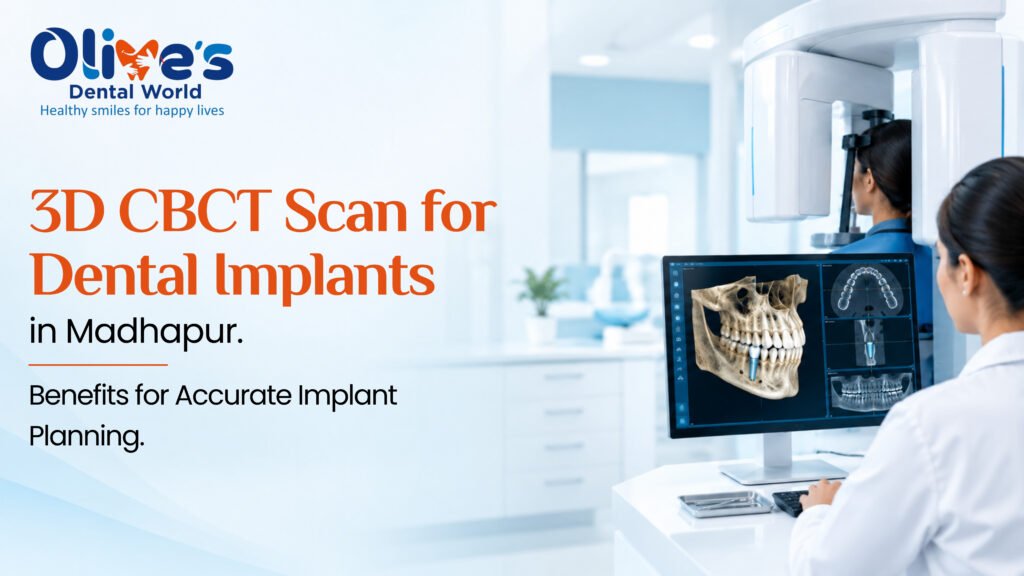
Dental implants are a reliable and popular solution for replacing missing teeth, restoring chewing ability, and improving the appearance of your smile. However, successful implant treatment requires careful planning, accurate measurements, and a thorough understanding of the patient’s jawbone and surrounding anatomical structures.
This is where a 3D CBCT scan for dental implants in Madhapur plays an important role. Cone Beam Computed Tomography (CBCT) is an advanced dental imaging technology that provides detailed three-dimensional views of the teeth, jawbone, nerves, and surrounding structures. It helps dentists plan implant placement with greater precision and make informed decisions based on each patient’s unique oral anatomy.
If you are considering dental implants in Madhapur, understanding the benefits of CBCT imaging can help you make more informed decisions about your treatment.
What Is a 3D CBCT Scan in Dentistry?
A CBCT scan is a specialized imaging procedure that uses a cone-shaped X-ray beam to capture detailed images of the teeth, jawbone, and surrounding oral structures. Unlike conventional dental X-rays, which provide two-dimensional images, CBCT produces a three-dimensional view of the treatment area.
This advanced imaging technology allows dentists to examine bone height, width, and density-related characteristics, identify important anatomical structures, and assess potential challenges before beginning dental implant treatment.
For patients seeking dental implants in Madhapur, a CBCT scan can provide valuable diagnostic information that supports personalized treatment planning.
Why Is CBCT Imaging Important for Dental Implant Planning?
Dental implant placement involves positioning a titanium implant within the jawbone to support a replacement tooth. For predictable treatment, the dentist needs to assess whether the available bone can accommodate the implant and determine the appropriate implant position.
A 3D CBCT scan provides information that may not be visible on routine dental X-rays.
Here are some of the key ways CBCT imaging supports implant planning.
1. Accurate Assessment of Jawbone Volume
Adequate jawbone volume is essential for supporting a dental implant. When a tooth has been missing for a long time, the surrounding bone may gradually shrink.
A CBCT scan helps the dentist measure the available bone height, width, and shape at the proposed implant site. These measurements help determine the appropriate implant dimensions and whether additional procedures, such as bone grafting, may be needed.
Benefit: Better understanding of the available bone helps the dentist develop a suitable treatment plan for the individual patient.
2. Identification of Nerves and Important Anatomical Structures
The jaw contains important structures, including nerves, blood vessels, and the maxillary sinuses. Their location must be considered carefully when planning dental implant placement.
In the lower jaw, the inferior alveolar nerve is particularly important because it provides sensation to parts of the lower lip and chin. In the upper jaw, the position of the maxillary sinus may influence implant placement.
CBCT imaging provides a three-dimensional view of these structures, helping dentists assess their proximity to the proposed implant site.
Benefit: Improved anatomical assessment supports safer treatment planning and helps reduce the risk of complications.
3. More Precise Implant Positioning
The position and angle of a dental implant influence its functional performance, the appearance of the final restoration, and the way chewing forces are distributed.
Using CBCT images, dentists can evaluate the available bone and plan the implant’s position in relation to neighbouring teeth, nerves, and other anatomical landmarks.
When appropriate, CBCT data can also be used with digital implant-planning software and surgical guide technology.
Benefit: Detailed preoperative planning can help the dentist position the implant in a way that supports the intended final tooth restoration.
4. Early Identification of Potential Complications
Every dental implant case is different. Some patients may have reduced bone volume, an unfavourable sinus position, or other anatomical factors that need special consideration.
A CBCT scan helps the dentist identify these factors before surgery rather than relying only on information obtained during the procedure.
This information may influence the choice of implant size, the surgical approach, the need for preparatory treatment, or whether a different treatment option should be considered.
Benefit: Identifying potential challenges in advance allows the dentist to prepare a more appropriate treatment plan.
5. Improved Planning for Bone Grafting and Sinus Augmentation
Patients who have experienced bone loss may require additional procedures before or during implant placement.
For example, patients with insufficient bone in the upper back jaw may be considered for sinus augmentation, while those with inadequate bone volume in other areas may benefit from bone grafting.
CBCT imaging helps the dentist evaluate the available bone and assess the anatomical conditions relevant to these procedures.
Benefit: Three-dimensional information supports better planning for complex implant cases and helps determine whether additional treatment may be necessary.
6. Support for Digital Implant Planning
Modern implant dentistry increasingly uses digital technologies to improve treatment planning and communication.
CBCT data can be combined with digital impressions or intraoral scans to create a more comprehensive representation of the patient’s teeth and jaw. Compatible software may allow the dentist to simulate implant placement and plan the final restoration before surgery.
In selected cases, this digital workflow can also support the design of a surgical guide to help transfer the planned implant position to the clinical procedure.
Benefit: Digital planning can improve coordination between the surgical and restorative stages of implant treatment.
CBCT Scan vs. Conventional Dental X-Ray
Understanding the difference between these imaging methods can help patients appreciate why a dentist may recommend a CBCT scan before implant surgery.
| Feature | Conventional dental X-ray | 3D CBCT scan |
|---|---|---|
| Image format | Two-dimensional | Three-dimensional |
| Bone assessment | Limited to the projected image | Cross-sectional assessment of bone dimensions |
| Anatomical structures | May overlap in the image | Provides improved spatial visualization |
| Implant planning | Useful for initial assessment in selected cases | Offers detailed information for complex implant planning |
| Radiation exposure | Generally lower, depending on the examination | Generally higher than routine intraoral dental X-rays |
The choice of imaging depends on the patient’s clinical needs. A CBCT scan is not necessary for every dental implant case, but it can be particularly valuable when three-dimensional information is needed to make treatment decisions.
Is a 3D CBCT Scan Necessary Before Every Dental Implant?
Not every patient requires CBCT imaging. The dentist will recommend the appropriate diagnostic imaging based on the medical and dental history, clinical examination, implant site, and complexity of the planned procedure.
A CBCT scan may be particularly useful when:
- The available jawbone volume is uncertain.
- The implant site is close to important nerves or the maxillary sinus.
- Bone grafting or sinus augmentation is being considered.
- Multiple implants or full-arch rehabilitation are planned.
- Conventional imaging does not provide sufficient information for safe and effective planning.
Because CBCT uses ionizing radiation, the scan should be prescribed when its expected diagnostic benefits outweigh the risks. The smallest suitable field of view and an appropriate imaging protocol should be used whenever possible.
What Happens During a Dental CBCT Scan?
For patients visiting a dental clinic in Madhapur, the CBCT scanning process is generally quick and straightforward.
Step 1: Initial consultation
The dentist examines your teeth and gums, reviews your dental history, and determines whether three-dimensional imaging is appropriate.
Step 2: Positioning
You will be asked to stand or sit in the designated scanning unit. The dental team will help position your head correctly to obtain clear images.
Step 3: Image capture
The CBCT machine rotates around your head and captures multiple images. The process is usually painless and does not involve injections or surgical procedures.
Step 4: Image analysis
The dentist reviews the reconstructed three-dimensional images to evaluate the jawbone, identify important anatomical structures, and assess the proposed implant site.
Step 5: Personalized treatment planning
Based on the scan findings and clinical examination, the dentist discusses the recommended implant approach, possible additional procedures, treatment timeline, and estimated cost.
Who Can Benefit from a 3D CBCT Scan in Madhapur?
CBCT imaging may be recommended for patients who:
- Have one or more missing teeth and are considering dental implants.
- Have experienced jawbone loss after tooth extraction.
- Need multiple implants or full-mouth implant rehabilitation.
- May require bone grafting or sinus augmentation.
- Have complex anatomical conditions that require detailed assessment.
Patients with diabetes, a history of gum disease, or other health conditions may require additional evaluation before implant treatment. A CBCT scan provides anatomical information, but it does not replace a comprehensive clinical examination or assessment of overall health.
How Does CBCT Contribute to Better Dental Implant Outcomes?
Successful dental implant treatment depends on several factors, including adequate bone support, healthy gums, proper implant positioning, good oral hygiene, and appropriate follow-up care.
CBCT imaging contributes by providing detailed information about the jawbone and surrounding structures before surgery. This can help the dentist select a suitable treatment approach, anticipate anatomical challenges, and coordinate implant placement with the final restoration.
However, a CBCT scan alone cannot guarantee implant success. Long-term outcomes also depend on the patient’s health, the quality of treatment, healing, and ongoing maintenance.
Choosing a Dental Clinic for CBCT and Dental Implants in Madhapur
If you are planning dental implant treatment, choosing a clinic that follows a comprehensive diagnostic and treatment-planning process is important.
Before proceeding, consider asking the dental team:
- Is CBCT imaging clinically necessary for my case?
- Will the scan provide sufficient information about my available jawbone?
- Do I need bone grafting or any other preparatory procedure?
- How will the implant position be planned in relation to the final tooth?
- What are the expected treatment stages, costs, and follow-up requirements?
A qualified dental implant specialist can explain the scan findings in understandable terms and help you compare suitable treatment options.
Frequently Asked Questions (FAQs)
1. What is the purpose of a CBCT scan before dental implant surgery?
A CBCT scan provides three-dimensional information about the jawbone and surrounding structures. It helps dentists evaluate bone dimensions, locate important anatomical landmarks, and plan implant placement.
2. Is a 3D CBCT scan painful?
No. The scan is non-invasive and painless. You simply remain still while the machine captures the required images.
3. How long does a dental CBCT scan take?
Image acquisition typically takes less than a minute, although the exact time varies by machine and scanning protocol. The complete appointment may take longer because of positioning and other clinic procedures.
4. Is CBCT imaging safe for dental implant planning?
CBCT uses ionizing radiation, so it should be prescribed only when clinically justified. The dentist should use an appropriate protocol that limits exposure while obtaining the information needed for treatment planning.
5. Can a CBCT scan determine whether I need bone grafting?
It helps the dentist assess bone volume and identify anatomical factors that may influence the need for bone grafting. The final decision also depends on the clinical examination and overall treatment plan.
6. Is a CBCT scan required for all dental implants in Madhapur?
No. The need for CBCT imaging depends on the individual case. Your dentist will recommend it when three-dimensional information is expected to improve diagnosis or treatment planning.
Plan Dental Implants with 3D CBCT Imaging in Madhapur
A 3D CBCT scan for dental implants in Madhapur is a valuable diagnostic tool that helps dentists evaluate jawbone dimensions, visualize important anatomical structures, and plan implant placement with greater precision. It can be particularly useful in complex cases, when bone grafting is being considered, or when conventional dental X-rays do not provide enough information.
If you are considering dental implants, consult an experienced dental professional to determine whether CBCT imaging is appropriate for your needs. A personalized treatment plan based on careful examination and suitable diagnostic imaging can help support safe, predictable, and long-lasting implant care.
Looking for dental implant treatment in Madhapur? Schedule a consultation with a qualified dental professional to discuss your options and find out whether a 3D CBCT scan can help plan your treatment.
contact :91 954 20 32 32
visit out website:https://olivesdentalworld.com/